
Twenty-five people from Denmark flew to the Canary Islands for a week of sunbathing, each with an ultraviolet meter strapped to one wrist. They kept diaries of what they wore and which sunscreen they put on. Every morning the same researcher examined each of them at seven places on the body, from the head down to the legs, and recorded any skin that had turned red. 1
All 25 of them got sunburned.
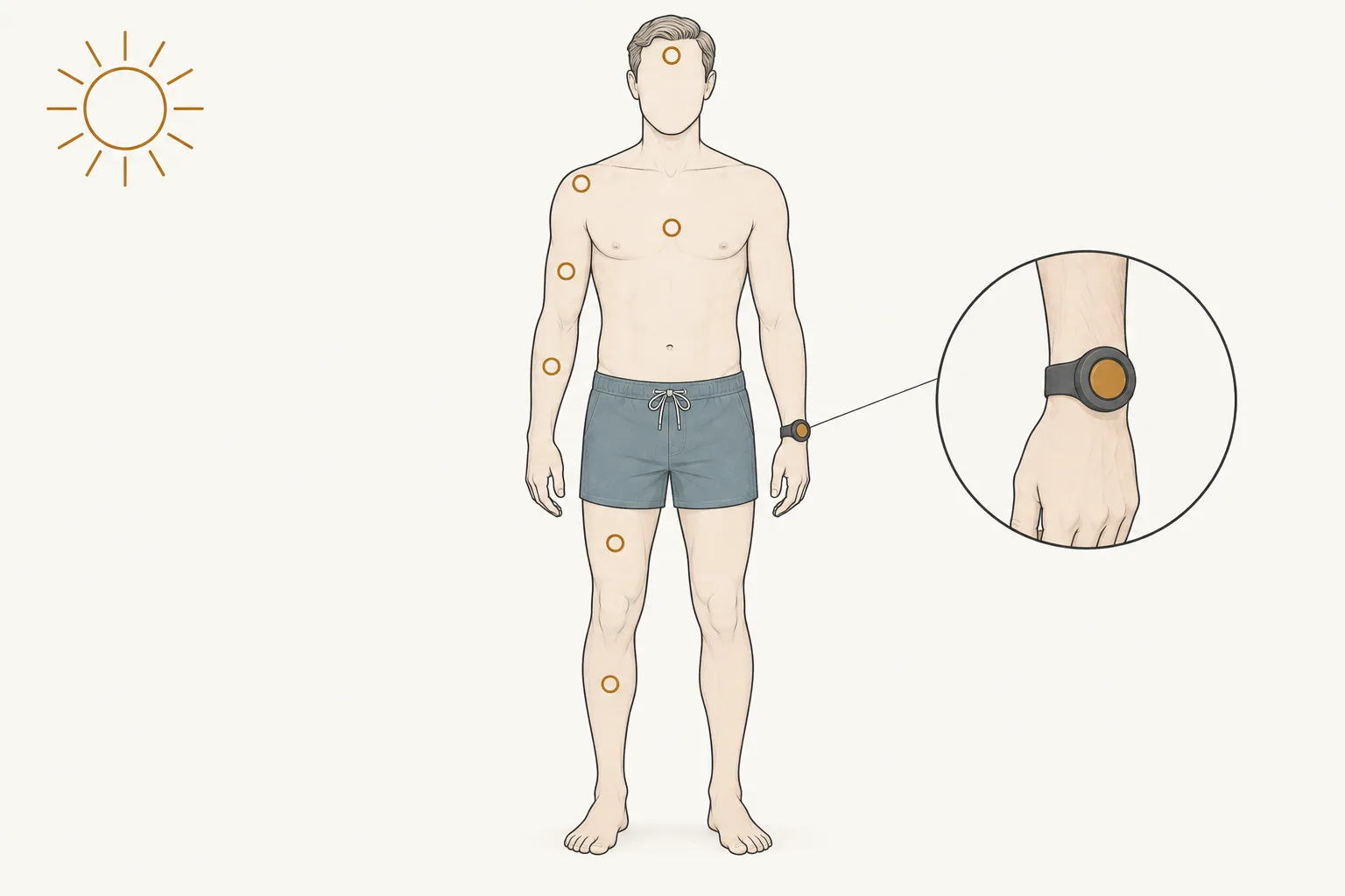
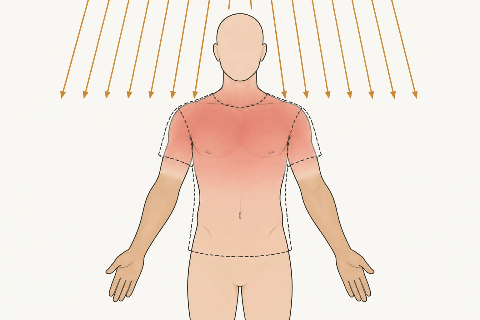
The meters counted ultraviolet in a unit called the standard erythema dose. Erythema is the medical word for redness of the skin, and one standard erythema dose is a fixed amount of the ultraviolet that reddens it. The week in the Canaries delivered 57 of those doses to each person on average. When the same Copenhagen research group had put meters on 164 people in Denmark going about their lives at home, the median for a whole year was 166 doses. A third of a year of sun had landed in seven days, much of it on chests and backs that are covered for most of a Danish year. Forearms, which see daylight all year, took the same dose as chests and burned less often. This is one of the few studies on this page that was run under the real sun. Most of the others used lamps. 1; 2
Since 1982, study after study has found that people who remember more sunburns turn up more often among melanoma patients, and most advice about the sun rests on that finding. This page covers what a sunburn is, what a tan is worth, how strong the tie to melanoma is, and where its best-known numbers came from.
What a sunburn is
In 1998 a team at St John's Institute of Dermatology in London had forty pale volunteers lie under a lamp that gave out one narrow wavelength of ultraviolet at a time. For each wavelength they found the smallest dose that left a patch of skin just visibly red a day later. That dose is called the minimal erythema dose, or MED. It is each person's own burn threshold, and most sunburn research is measured in it. Then the team gave fresh patches of skin half, one, two, and three times the MED. Within five minutes they cut out a plug of skin 4 millimeters across and stained it to count one kind of DNA damage, called a pyrimidine dimer. 3
A pyrimidine dimer is the commonest injury ultraviolet does to DNA. Two of the four DNA letters, thymine and cytosine, are called pyrimidines. Where two of them sit side by side on a strand, the light they absorb can weld them together, and the weld kinks the strand so that the cell can no longer read or copy it properly.
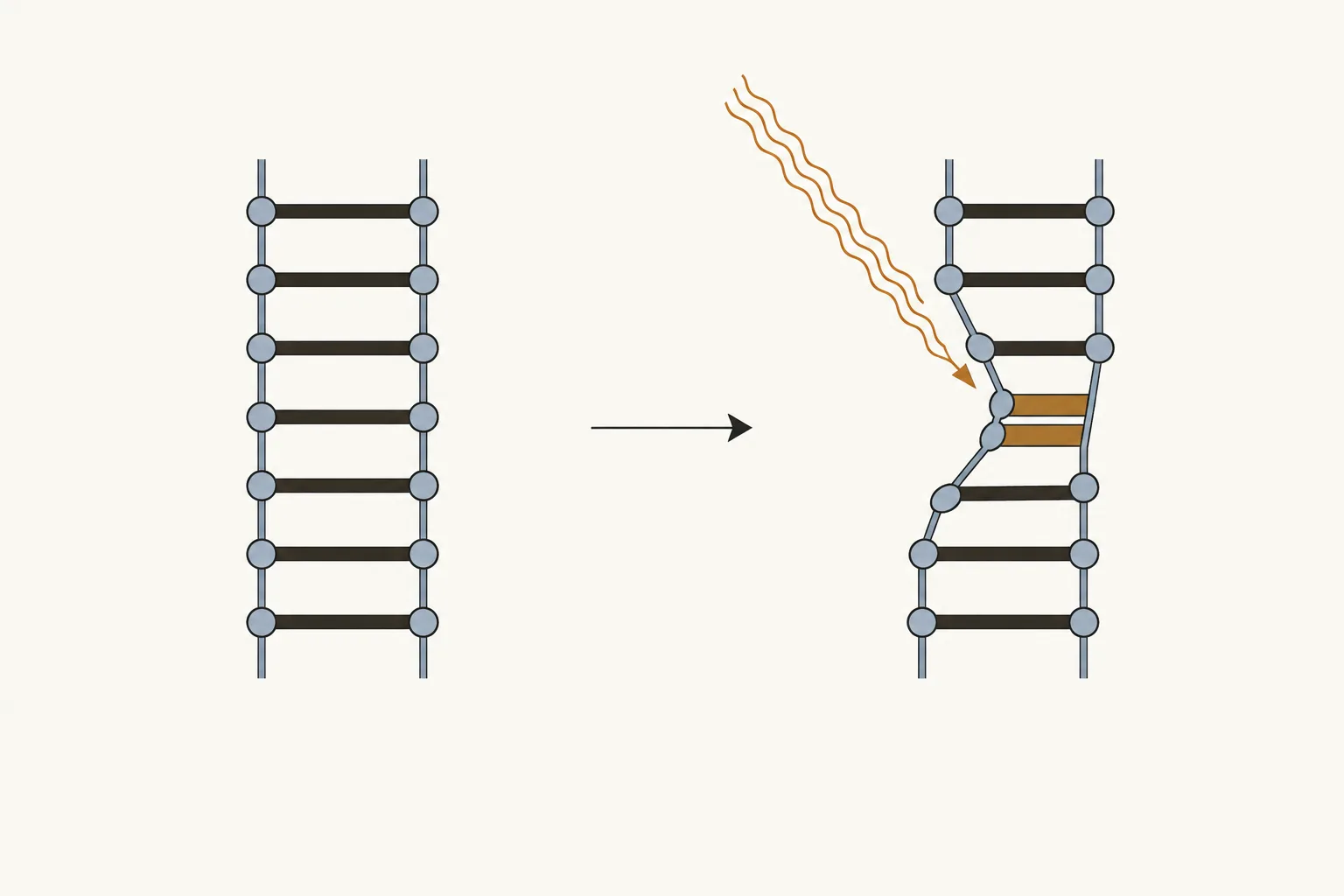
The London team drew two curves against wavelength, one for how easily the skin reddened and one for how many dimers it held. From 280 to 340 nanometers, which covers the burning part of sunlight, the two curves lay almost on top of each other. Whatever molecule absorbs the light that starts a sunburn, it absorbs exactly like DNA. The team concluded that it mostly is DNA. 3
Dimers form while the light is still on, and the biopsies taken at five minutes were already full of them. The redness takes hours, and in 2009 Lesley Rhodes's group in Manchester watched it arrive. Thirty-two volunteers took four times their own MED from a UVB lamp on a patch of skin, and for three days the researchers raised small suction blisters over it and drew off the fluid. Redness showed by 4 hours and peaked between 18 and 24. It rose and fell with the prostaglandins, chemical messengers that widen blood vessels, and the extra blood is where the red and the heat of a sunburn come from. Aspirin and ibuprofen work by blocking the same messengers. Neutrophils, the white blood cells that arrive first at any injury, crowded into the skin at 24 hours and had left by 72. By the third day the skin was making more and more of a different messenger, one that shuts inflammation down. A sunburn ran in that order in every volunteer: the blood vessels opened, the white cells came and went, and the skin shut the reaction down. 4
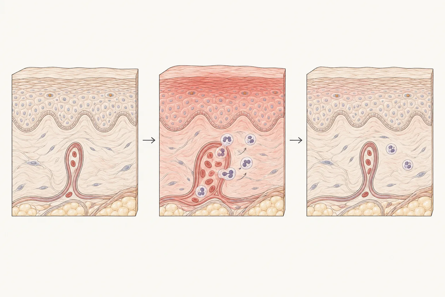
A sunburn, then, is the cleanup, and it runs a day behind the injury. Skin cells carrying too many dimers are told to die by p53, a protein that checks DNA for damage, and the next day they show under the microscope as shrunken dark bodies that pathologists call sunburn cells. The damaged cells also spill fragments of their own RNA. A San Diego laboratory found in 2012 that neighboring cells pick those fragments up with a receptor they otherwise use to detect viruses, and that mice bred without the receptor did not raise the usual inflammatory alarm after a burning dose. 5; 6
How much light it takes to burn differs a great deal between people. A dermatology clinic in Grenoble spent a winter finding out, and published the result in 1982. Its 303 volunteers, aged 5 to 89, lay under a 2,500-watt xenon lamp while a motor slid a plate across nine openings, so that nine neighboring patches of each back got nine rising doses. Volunteers with light complexions reddened at an average of 658 millijoules per square centimeter and those with dark complexions at 1,607. All 303 volunteers were white, so light and dark here are shades of white skin, and the burn threshold still differed by a factor of two and a half. Once complexion was known, hair and eye color added nothing. 7
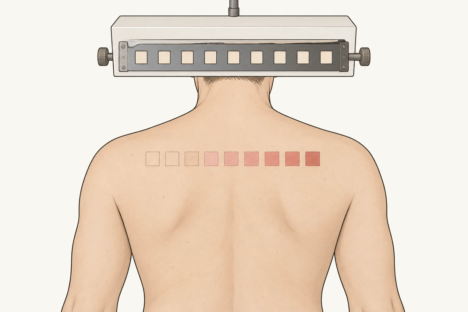
Across the whole human range the spread is wider. In a 2018 Manchester study the burn threshold ran about sevenfold from the palest volunteers to the darkest. The familiar six skin types, from type I, which always burns and never tans, to type VI, were drawn up by the Boston dermatologist Thomas Fitzpatrick in 1975 so that clinics could pick a starting dose for psoriasis patients under ultraviolet lamps. They rest on what people say about their own skin. 8; 9
That Manchester study was built to see what happens below the burn threshold. Thirty-nine volunteers of every skin type stood in a lamp cabinet four times, a month apart, and took a fifth, then two fifths, three fifths, and four fifths of their own MED. Even the smallest dose left dimers in the skin of every volunteer. Two days later most had been repaired. The team found no dose too small to leave some, and a Lausanne group had shown the same thing twenty years earlier, along with p53 switched on through every layer of the epidermis. Redness is a late and coarse gauge of what has happened to the DNA underneath. 8; 5
A tan begins with DNA damage and protects about as well as SPF 2
Melanocytes, the pigment cells, sit at the base of the epidermis and hand packets of melanin to the ordinary skin cells around them. What tells them to make more was worked out in David Fisher's laboratory at Dana-Farber in Boston in 2007. Ultraviolet damages the DNA of the ordinary skin cells. In those cells p53, the same protein that tells the worst-hit cells to die, switches on a gene called POMC. The POMC protein is cut into pieces, and one piece is the hormone that tells melanocytes to make melanin. Mice without p53 could not tan. 10
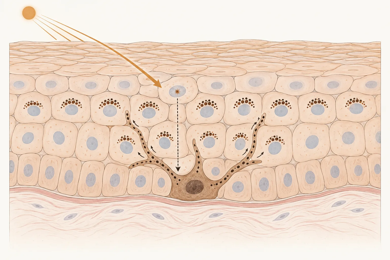
Another piece cut from the same protein is beta-endorphin, one of the body's own opioids. Seven years later the same laboratory gave shaved mice a small dose of ultraviolet day after day for weeks, and the endorphin in their blood rose. Then the researchers injected naloxone, the drug paramedics carry to reverse a heroin overdose. The mice shook and trembled like animals coming off an opiate, and afterward they avoided the box where it had happened. Mice bred without beta-endorphin did none of this. Nobody has run the equivalent test in people. 11
UVA and UVB tan skin in different ways. UVA is the longer-wave ultraviolet, most of what reaches the ground and most of what a tanning bed puts out, and it darkens melanin that is already in the skin, within minutes. UVB, the shorter burning band, makes melanocytes build new melanin over several days. In 2011 researchers from the US National Cancer Institute and the skincare company Beiersdorf tanned patches of volunteers' backs for two weeks with UVA lamps, UVB lamps, or both, until the patches looked alike. Then they hit every patch with the same test dose and counted the DNA damage. The UVB tans gave modest protection. The UVA tans gave essentially none. 12
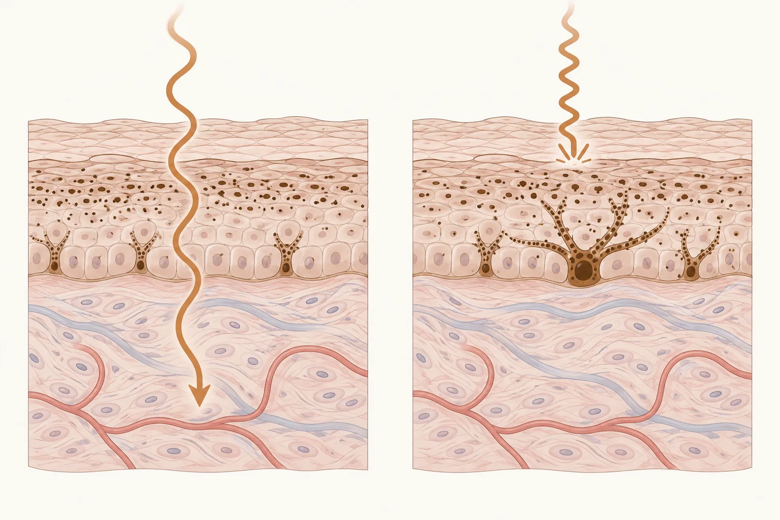
In 1998 the London group gave sixteen volunteers a lamp dose below their burn threshold every day for two weeks, waited a week, and measured how much more light the tanned skin could take before it reddened. It could take 1.4 to 2.3 times as much. About a fifth of that came from thickening of the dead outer layer of skin and had nothing to do with pigment. 13
In 2002 they ran it again with six people who tan poorly and six who tan well, and this time counted dimers too. Protection came out near 2 against redness and near 2 against DNA damage. It was the same in both groups, although the good tanners had gone much darker. The tan had a cost. Dimers built up in everyone's skin across the two weeks. A week after the last dose the good tanners had cleared 69 percent of theirs, and the poor tanners 37 percent. 14
A Copenhagen group built tans with lamps and followed them to their end. Building one slowly, out of many small doses, took three to four times the total ultraviolet of building it fast, and it faded in four to five months on every skin they tested. By the time someone from Denmark flies south in February, last summer's tan has gone. 15
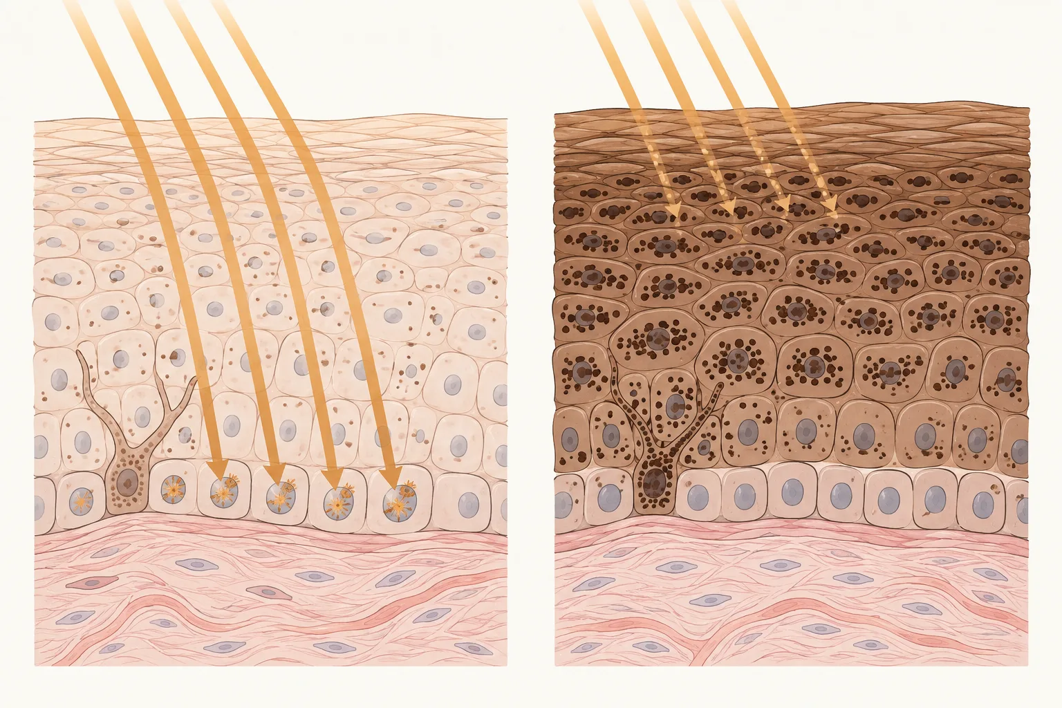
The pigment a person is born with is another matter. In 2018 the London group gave six very fair volunteers and six Black West Africans the same kind of lamp exposure and counted dimers layer by layer through the epidermis. In the bottom layer, where the dividing cells and the melanocytes live, the dark skin needed 59 times the dose to take the same damage as the fair skin. The two-week tans in the earlier London experiments had raised that dose about 2 times. 16; 14
People who recall more sunburns get more melanoma
By 1980 researchers knew that melanoma was commonest in fair-skinned people living in strong sun, and the first interview studies had found, to general surprise, that it did not rise with total hours outdoors. The melanoma page tells that story. Sunburn was the next thing to ask about.
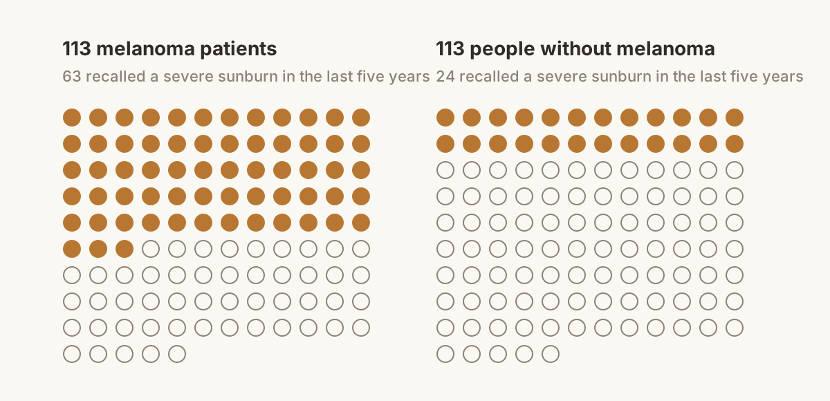
In Glasgow, Rona MacKie and Thomas Aitchison interviewed 113 melanoma patients and 113 people matched to them by age and sex. Sixty-three of the patients recalled a severe sunburn in the five years before diagnosis. So did 24 of the comparison group. A third of the patients had never left Britain. 17
In Boston a year later, one interviewer telephoned 111 melanoma patients from Massachusetts General Hospital and 107 friends those patients had nominated. More than half the patients recalled a blistering sunburn in adolescence. Fewer than a third of the friends did, which makes the odds about 2 to 1. Having been a poor tanner as a teenager also carried odds of about 2. No measure of total lifetime sun separated patients from friends. One of the study's five authors was Thomas Fitzpatrick, the dermatologist who had drawn up the six skin types. 18
In Queensland in 1985 Adele Green's group spelled out the logic. Nobody could measure the ultraviolet that had reached a person's melanocytes decades earlier, so they let the burn stand in for the meter. A sunburn that hurt for more than two days proved that a heavy dose had got through, whatever the person's coloring. One interviewer counted such burns, decade by decade of life, for 183 melanoma patients and 183 neighbors drawn from the electoral roll. After adjusting for the number of moles on the arms, people with two to five severe burns had 1.5 times the melanoma risk of people with none or one. People with six or more had 2.4 times. 19
The Queensland team also tested whether cancer patients simply search their memories harder. Their patients with lentigo maligna, the slow melanoma of old sun-weathered faces that nobody had connected to sunburn, recalled about as many burns as the neighbors did. Only the patients with the common kinds of melanoma recalled more. If a cancer diagnosis by itself made people remember extra sunburns, the lentigo maligna patients would have remembered extra too. 19
Two sentences found on many cancer charity websites grew out of studies like these. The first is "One blistering sunburn in childhood or adolescence more than doubles your chances of developing melanoma later in life." Its source is the Boston telephone study of 111 patients and their friends.
The second is "Your risk for melanoma doubles if you have had more than five sunburns." The Skin Cancer Foundation's footnote for it leads to a 2001 study from Erlangen in Germany, which interviewed 603 patients and 627 comparison people in seven European countries. Its authors were testing whether burns in childhood are worse than burns later on. They found the same climb in risk for burns before age 15 as after, and put the finding in their title: epidemiology does not support the existence of a critical period. 18; 20; 21
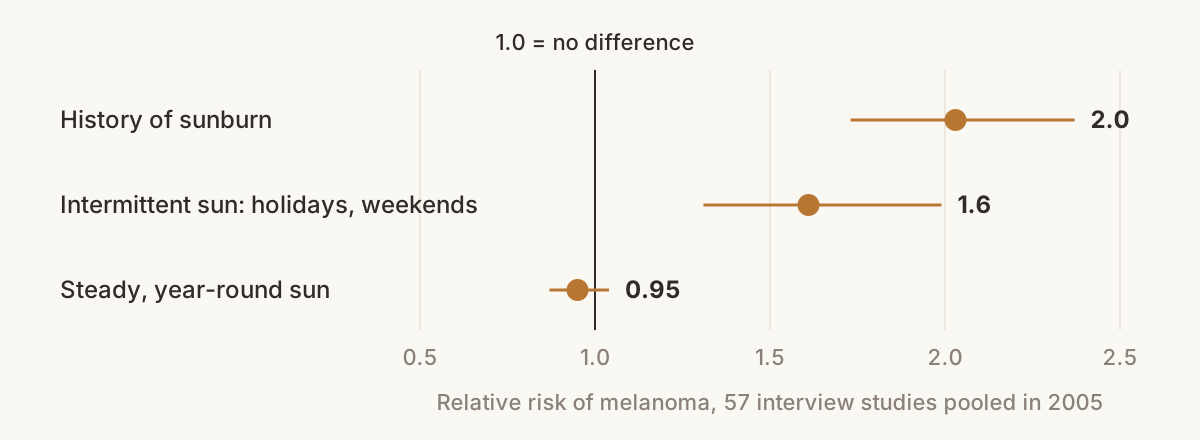
By 2005 there were 57 interview studies to pool. What each of them had observed was that melanoma patients recalled sunburns more often than the people they were compared with. Pooling turns those differences into a relative risk, a number where 1.0 means the two groups got melanoma equally often and 2.0 means one group got it twice as often. People with a history of sunburn came out at about 2.0. People with a lot of intermittent sun, the holiday and weekend kind, came out at 1.6. People with steady, year-round exposure came out at 0.95, which is no difference. 22
Every one of those studies asked people about their burns after the diagnosis. Two long-running American health studies asked before. In the 1980s and early 1990s, 87,166 nurses and 32,959 male health professionals described their sunburn histories while none of them had melanoma, and researchers then followed them until 2010. The same questionnaires recorded hair color, moles on the arms, how their skin had reacted to sun in childhood, family history of melanoma, and hours spent in summer sun, and the analysis adjusted for all of these. Women who had ever had a severe sunburn were diagnosed with melanoma at 1.63 times the rate of women who never had, and men at 2.41 times. Burns on the trunk counted for more than burns on the face, arms, or legs. 23
Almost everyone in those two studies had been severely sunburned at some point. Only 47 of the 774 women who developed melanoma said they never had, and only 11 of the 391 men. Those few people are the group everyone else is compared with, so the 1.63 and the 2.41 rest on small numbers. Among the women, more burns also did not mean more risk. One to five burns carried 1.51 times the rate of the never-burned, and six to ten burns carried 1.44 times. 23
A Norwegian study reported in 2022 on 169,768 women who had filled in questionnaires about their natural coloring, sunbathing holidays, tanning bed use, and how often they burned in childhood, adolescence, and adulthood. The researchers sorted the women by the shape of their sunburn history across life. Women who had burned often at every age were diagnosed with melanoma at 1.50 times the rate of women who had rarely burned. Women who burned often as girls and then stopped came in at 1.44. 24
Whether childhood burns count for more is unsettled
That last Norwegian figure is one piece of evidence that early burns leave a lasting mark. There are others. British migrants who reached Australia before the age of ten grew up to get melanoma at the Australian rate, while those who arrived after fifteen got about a quarter of it. A 2025 analysis of 44,021 American radiologic technologists found melanoma risk rising about 3 percent with each blistering burn before age 15 and no detectable rise for burns after. And in 2001 Frances Noonan's group gave a single burning dose of ultraviolet to mice engineered to be melanoma-prone. Newborn mice that got the dose went on to develop melanomas. Adult mice that got it did not. 24; 25; 26; 27
Against that stands the Erlangen study, with its equal risks before and after 15, and the largest pooling of burn studies, from 2008. Across 51 study populations, melanoma risk rose with the number of burns in childhood, in adolescence, and in adulthood. When the authors put the three periods on one scale, five burns per decade, the adult burns carried the largest risk. The Erlangen authors wrote that "the hazardous impact of sunburns seems to persist lifelong." The 2008 authors did not claim that adult burns are worse. Childhood is a short stretch of life remembered from a long way off, and changing the scale changed the answer. 20; 28
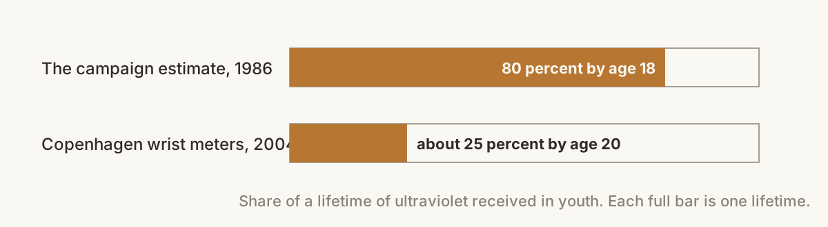
The case for childhood often arrives with the claim that 80 percent of a lifetime's sun lands before age 18. Sun-protection campaigns adopted that figure in 1986 as an estimate. The Copenhagen meters tested it. Annual dose had no relation to age, and only about 25 percent of a lifetime's dose had arrived by age 20. 2
A sunburn measures the skin as well as the sun
Using the burn as a meter has a catch. A burn records how much ultraviolet arrived. It also records how easily that person's skin burns, and the Grenoble thresholds differed by a factor of two and a half among white French volunteers alone. And it records how long it had been since that patch of skin last saw the sun, which is why the Danish holidaymakers' forearms burned less often than their chests.
So when a study finds that people who burned more get more melanoma, part of what it has found is that people with skin that burns get more melanoma. That was known before anyone counted burns. In Oslo in 1979, people who said they tolerated the sun poorly had more than twice the melanoma risk of people who tolerated it well, and in Boston the poor tanners had their doubled odds whether they recalled burns or not. Studies that adjust for skin type still find that burns count, by a smaller margin. No study has been able to pull the three apart cleanly. 29; 18; 28
The 0.95 for steady exposure fits the same picture. Skin that meets the sun daily thickens, darkens, and burns less at a given dose. Outdoor workers have skin like that, and in the pooled interview studies they had no more melanoma than indoor workers. A 2026 Danish registry study of nearly three million workers did find more melanoma with more years of outdoor work, so that question has reopened. The pattern that goes with melanoma most consistently is a large dose landing on skin that was not ready for it, in a person whose skin burns. 22; 30
In the nurses' cohort, about 40 of every 100,000 women who had ever burned were diagnosed with melanoma in a given year, against about 18 of every 100,000 who never had. Those are raw rates, before any adjustment. Nearly all of the ever-burned women, year after year, were not diagnosed with anything. No study of any design can tie one person's tumor to one afternoon. 23
Sunscreen's record at preventing burns on holiday is mixed, for a reason a French and Swiss trial caught in 1997. It handed 87 holidaymakers aged 18 to 24 an unlabeled sunscreen, SPF 10 or SPF 30. The two groups used the same amount and burned equally often. The SPF 30 group had stayed out longer, 72.6 hours of sunbathing against 58.2. The one randomized trial with a melanoma count is from Nambour in Queensland, where 1,621 adults were assigned to daily sunscreen or to their usual habits. Years later the daily group had 11 melanomas and the other group 22. That is half as many, but both counts are small, and small counts can differ by chance. By the trial's own statistics the true effect of daily sunscreen could be anywhere from a cut of three quarters in melanomas to no cut at all. 31; 32
Tanning beds raise melanoma risk by about a fifth, and by more with early or heavy use
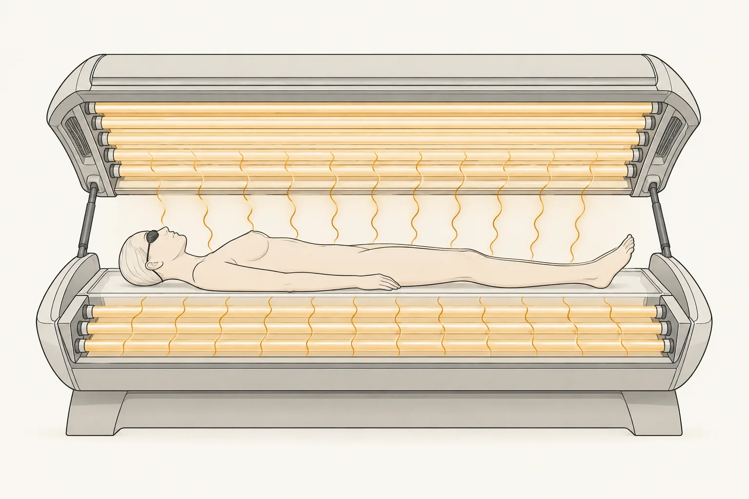
A tanning bed sells the dose on purpose, which ought to make it the easiest exposure to study. In 2006 a working group of the International Agency for Research on Cancer pooled the studies then available. People who had ever used a sunbed had 1.15 times the melanoma risk of people who never had. People who first used one before age 35 had 1.75 times. That second figure became "sunbeds raise your risk by 75 percent," and in 2009 the agency moved tanning devices into Group 1, its list of proven human carcinogens. 33
A 2012 update in the BMJ pooled 27 studies and put ever-use at 1.20. It put first use before 35 at 1.87. That December the journal printed a correction. A data file had been corrupted, and the true figure was 1.59. The 87 percent is still in circulation, on Cancer Research UK's news pages among other places. 34; 35; 36
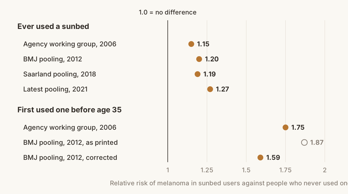
In 2018 a group at Saarland University in Germany pooled 31 studies, reached almost the same ever-use figure, 1.19, and drew the opposite conclusion. They graded every included study as poor in quality, mostly because people were asked about tanning after their diagnosis. The association disappeared when they looked only at European studies, only at studies after 1990, or only at the studies least open to bias. They called a causal link unproven. 37
Two studies built to answer those objections point the other way. In Minnesota, where indoor tanning was common, 63 percent of 1,167 melanoma patients had tanned indoors against 51 percent of 1,101 people drawn from driver's license lists. After adjusting for the known risk factors, the odds of melanoma were 1.74 times higher in those who had, and they rose with the years, the hours, and the number of sessions. In Norway, 141,045 women reported their tanning habits before anyone was ill. Over the next fourteen years the third who had tanned most had 1.32 times the melanoma risk of women who never had. Women who started before 30 had 1.31 times the risk, and their melanomas were diagnosed 2.2 years younger on average. The Norwegian analysis adjusted for sunburns and sunbathing holidays, because women who use sunbeds also do more of both. The latest pooling, of 36 studies in 2021, put ever-use at 1.27 and melanoma before age 50 at 1.75. 38; 39; 40
So the agreed number for ever having used a sunbed is an increase of roughly a fifth to a quarter, on a disease that about 2 in 100 Americans are diagnosed with in a lifetime. Starting young and going often raise it further. 40; 41 And since most of a sunbed's output is UVA, the tan it builds is the kind that protected nothing in the 2011 test. 12
What is still unknown
Nobody has shown why a week in the Canaries should matter more than a working life outdoors. One proposal is that a sudden load of dimers swamps the repair enzymes. Another is that a burn shuts down the skin's immune patrols for a few days. A third is that a big dose pushes melanocytes to divide while they still carry damage. Each has laboratory support, and no experiment in people has been able to choose between them.
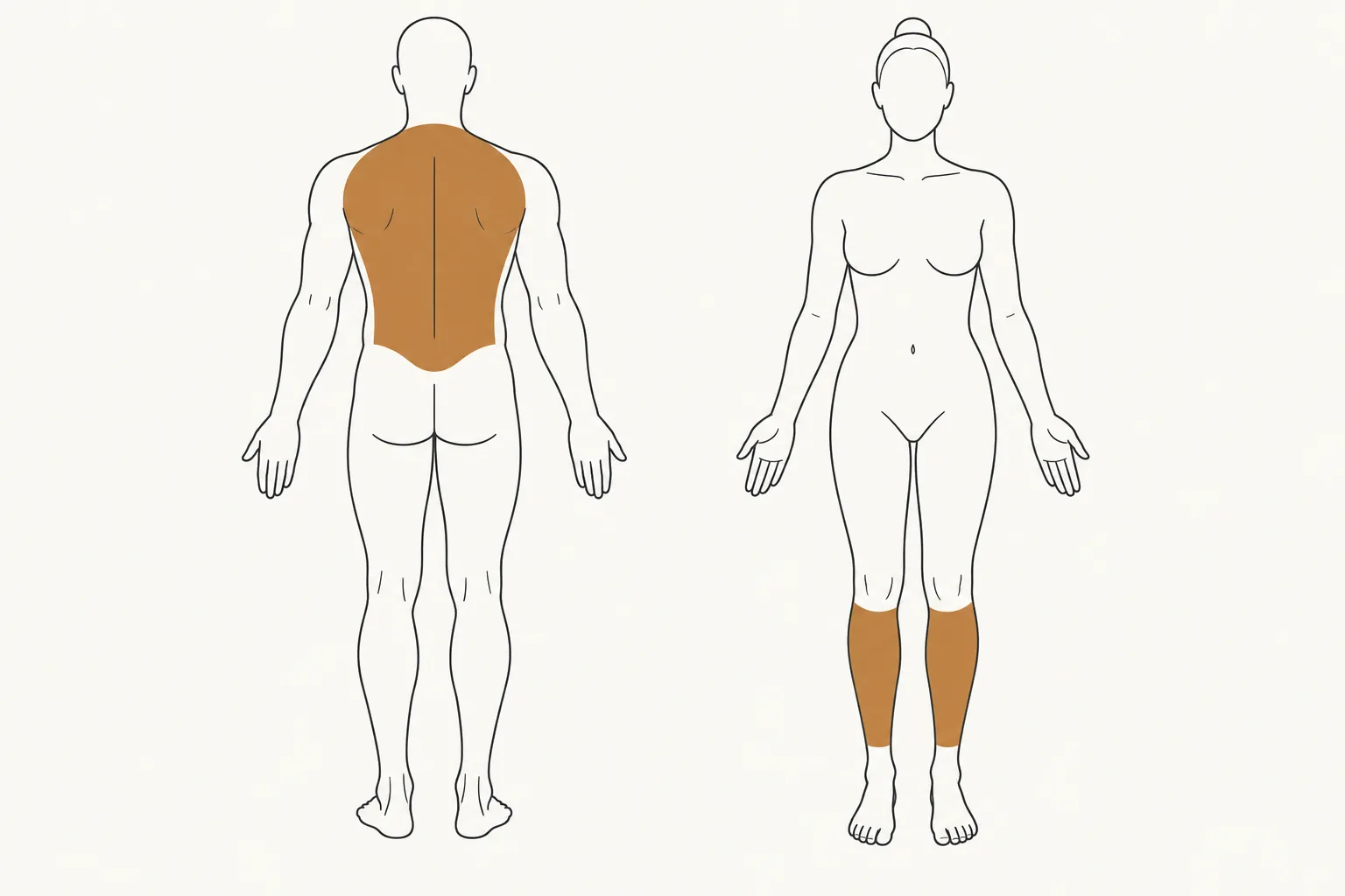
Melanoma favors the backs of men and the legs of women, skin that is bare a few weeks a year. No measure of dose in use, burns included, accounts for that map.
The Brisbane melanocyte trial gave 57 volunteers one dose of simulated sun, twice their own MED, on skin that had never been exposed. Two weeks later the number of melanocytes in that skin had roughly doubled. In carriers of the red-hair variants of the gene MC1R, the people most likely to get melanoma, the increase was smaller, 97 percent against 164. Sunscreen put on beforehand blocked the DNA damage, the p53 response, and the multiplying. Why the highest-risk melanocytes respond least has not been explained. 42
And none of the melanoma studies on this page measured anyone's sunlight. They asked people what they remembered. In the Canary Islands the wrist meters could see a burn coming: each extra standard erythema dose on one day raised the odds of new redness the next morning by about 11 percent. No study of melanoma has had a meter on anyone's wrist. 1
Key studies
- Young and colleagues, 1998, London. Redness and DNA dimers follow the same curve across wavelengths. 3
- Rhodes and colleagues, 2009, Manchester. The three-day chemistry of a sunburn. 4
- Amblard and colleagues, 1982, Grenoble. Burn thresholds in 303 volunteers aged 5 to 89. 7
- Shih and colleagues, 2018, Manchester. A fifth of a burn dose leaves DNA damage in every skin type. 8
- Cui and colleagues, 2007, Dana-Farber. p53 starts the tan. 10
- Fell and colleagues, 2014, Dana-Farber. Ultraviolet, beta-endorphin, and naloxone withdrawal in mice. 11
- Miyamura and colleagues, 2011. A UVA tan gives essentially no protection. 12
- Sheehan and colleagues, 1998 and 2002, London. A two-week tan is worth a factor of about 2. 13; 14
- Ravnbak and colleagues, 2008, Copenhagen. A tan fades in four to five months. 15
- Fajuyigbe and colleagues, 2018, London. Deeply pigmented skin protects its bottom layer 59-fold. 16
- MacKie and Aitchison, 1982, Glasgow. Severe sunburn in the five years before diagnosis. 17
- Lew and colleagues, 1983, Boston. Blistering adolescent burns; the source of the one-burn sentence. 18
- Green and colleagues, 1985, Queensland. The burn as a meter, with a check on patients' memories. 19
- Pfahlberg and colleagues, 2001, Erlangen. More than five burns doubled risk, before age 15 and after alike. 20
- Gandini and colleagues, 2005. Fifty-seven studies pooled. 22
- Dennis and colleagues, 2008. Fifty-one populations; burns counted at every age. 28
- Wu and colleagues, 2016. Burn histories collected before diagnosis in two US cohorts. 23
- Lergenmuller and colleagues, 2022, Norway. Lifetime sunburn histories in 169,768 women. 24
- Noonan and colleagues, 2001. One burning dose gave newborn mice melanoma, and adults none. 27
- Thieden and colleagues, 2004, Copenhagen. A quarter of lifetime ultraviolet arrives by age 20. 2
- Petersen and colleagues, 2013, Copenhagen. Twenty-five sunbathers, twenty-five sunburns. 1
- Autier and colleagues, 1999. Higher SPF, longer sunbathing, the same burns. 31
- Boniol and colleagues, 2012, with its correction. Sunbed use before 35: 1.59. 34; 35
- Burgard and colleagues, 2018, Saarland. The case that the sunbed evidence is weak. 37
- Lazovich and colleagues, 2010, Minnesota. Indoor tanning in 1,167 patients. 38
- Ghiasvand and colleagues, 2017, Norway. Tanning habits recorded before diagnosis in 141,045 women. 39
- Hacker and colleagues, 2013, Brisbane. Melanocytes doubled after one exposure, least in MC1R carriers. 42
Definitions
- Cohort study. A study that questions a large group of healthy people first and then follows them for years to see who becomes ill. The answers cannot be colored by a diagnosis, because nobody has one yet. The American nurses' study and the two Norwegian studies on this page are cohort studies.
- Epidermis. The outer layer of the skin, about a tenth of a millimeter thick over most of the body. Its top is a sheet of dead cells. Its bottom layer holds the dividing cells and the melanocytes.
- Erythema. The medical word for redness of the skin.
- Fitzpatrick skin type. A six-step scale of how skin responds to sun, from type I, which always burns and never tans, to type VI, which is deeply pigmented and almost never burns. People are placed on it by their own account of their skin.
- Interview study. Called a case-control study in the journals. Researchers interview people who already have a disease and a comparison group who do not, and look for differences in what the two groups recall.
- Lentigo maligna. A slow-growing kind of melanoma found mostly on the faces of older people with many years of sun behind them.
- MC1R. A gene that helps set which kind of melanin a person makes. Some variants of it give red hair, freckles, and skin that burns easily.
- Melanin. The brown-black pigment of skin and hair. It absorbs ultraviolet.
- Melanocyte. The cell that makes melanin. Melanocytes sit in the bottom layer of the epidermis and pass their pigment to the cells around them. Melanoma is the cancer of this cell.
- Minimal erythema dose (MED). The smallest dose of ultraviolet that leaves one person's skin just visibly red a day later. It is that person's own burn threshold, and it differs severalfold between people.
- Neutrophil. The white blood cell that arrives first at an injury.
- p53. A protein that checks a cell's DNA for damage. It can pause the cell for repairs or order it to die. In skin it also starts the tanning signal.
- POMC. A gene whose protein is cut into several smaller hormones. One of them tells melanocytes to make melanin. Another is beta-endorphin, one of the body's own opioids.
- Pooled analysis. Called a meta-analysis in the journals. It combines the results of many separate studies into one estimate.
- Prostaglandins. Chemical messengers that widen blood vessels and cause the redness, heat, and soreness of inflammation. Aspirin and ibuprofen block them.
- Pyrimidine dimer. Two neighboring DNA letters welded together by ultraviolet. It is the commonest injury ultraviolet does to DNA.
- Randomized trial. A study in which chance decides who gets the treatment, so that the groups are alike in everything else, apart from chance.
- Recall bias. The error that comes in when people who are ill remember their past differently from people who are well.
- Relative risk. How often one group gets a disease, divided by how often another group gets it. 1.0 means no difference, 2.0 means twice as often, and 0.5 means half as often. Odds ratios and hazard ratios are close relatives and read the same way.
- SPF. Sun protection factor. How many times more ultraviolet it takes to redden skin through a product, tested in a laboratory with a thick, even layer.
- Standard erythema dose (SED). A fixed amount of skin-reddening ultraviolet, the same for everyone, used to report what a meter has measured.
- Sunburn cell. A skin cell that has been told to die after ultraviolet damage. It shows under the microscope as a shrunken dark body.
- UVA. Ultraviolet with wavelengths from 315 to 400 nanometers. It is most of the ultraviolet that reaches the ground and most of what a tanning bed gives out.
- UVB. Ultraviolet with wavelengths from 280 to 315 nanometers. It is a small part of sunlight and does most of the burning.
Statistics
In 40 pale London volunteers under single-wavelength lamps, the median burn threshold was 0.025 joules per square centimeter at 300 nanometers and 32.0 at 360, about 1,280 times more light for the same redness. 3
Light at 280 nanometers left almost 40 times less DNA damage in the deepest layer of the epidermis than in the top layer, while light from 300 to 360 nanometers damaged every layer alike. 3
Across 39 Manchester volunteers of skin types I to VI, the mean burn threshold rose from 21 to 152 millijoules per square centimeter, which is 2.1 to 15.2 standard erythema doses. 8
Every extra fifth of a burn dose added 1.6 nanomoles per liter of 25-hydroxyvitamin D, with a 95 percent interval of 1.1 to 2.0, and skin type made no difference. 8
In Manchester volunteers of skin types IV to VI, DNA damage in the bottom layer of the epidermis stayed undetectable across the whole range from a fifth to four fifths of a burn dose, while vitamin D still rose. 8
A solar simulator dose too small to redden the skin raised p53 to about 41 stained cells in every 1,000 epidermal cells, up from fewer than 10. 5
After a solar simulator dose below the redness threshold, roughly 10 to 25 percent of the volunteers' epidermal cells stained for pyrimidine dimers. 5
The mean burn threshold of 303 Grenoble volunteers under a xenon lamp was 889 millijoules per square centimeter, and 97.5 percent of them needed more than 350. 7
Burn thresholds in Grenoble fell with age, from a mean 1,184 millijoules per square centimeter in volunteers aged 5 to 15 down to 818 in those over 60. 7
The 26 freckled Grenoble volunteers reddened at a mean 748 millijoules per square centimeter against 1,009 for the 277 without freckles. 7
After four times their own burn dose from a UVB lamp, the Manchester volunteers' redness reading rose from 39.9 to 97.2 within 4 hours, peaked at 171.1 by 24 hours, and was still 156.6 at 72 hours. 4
Neutrophils in the dermis of burned Manchester skin rose from 3.07 per microscope field to 8.6 at 24 hours and were back to baseline by 72 hours. 4
Two weeks of daily lamp doses at half the burn threshold raised the threshold 1.4 times in skin type II volunteers, and doses at three quarters of it raised the threshold 2.1 times. 13
A two-week lamp tan in 12 London volunteers gave protection factors in the region of 2 to 3 against both redness and thymine dimers, and skin type IV gained no more than skin type II. 14
Before any tanning, the untanned burn threshold of the skin type IV volunteers was 1.8 times that of the skin type II volunteers. 14
Splitting a tanning course into 6 or 12 lamp sessions cut the dose per session to a half and a third, but raised the total ultraviolet needed by 3 and 4 times, in 12 Scandinavian and 12 South Asian volunteers. 15
Taken across the whole epidermis rather than one layer, the DNA protection factor of deeply pigmented skin over very fair skin was 8.0, with a 95 percent interval of 6.7 to 9.9. 16
One day after a dose of simulated sun at twice the burn threshold, 25 percent of the Brisbane volunteers' skin cells and 36.6 percent of their melanocytes carried pyrimidine dimers, against none in the unexposed and sunscreened patches. 42
p53 was present in 1 percent of the Brisbane volunteers' ordinary skin cells at baseline, rose to 15.1 percent a day after the dose, and had fallen back to 2.7 percent at 14 days. 42
In 16 fair-skinned London volunteers given twice their burn dose, thymine dimers cleared with a half-life of 33.3 hours and lesions were still visible seven days later. 43
The other main ultraviolet lesion, the 6-4 photoproduct, cleared from the same volunteers' skin with a half-life of 2.3 hours, about 14 times faster than thymine dimers. 43
Three to four days after a burning dose, the number of cells copying their DNA in that skin had risen up to 4.7 times. 43
In 17 Dublin volunteers, dying cells first appeared 24 hours after a solar simulator dose, peaked at 48 hours at 4.22 cells per section, and had fallen to 1.61 by 72 hours. 44
At the dose that makes the first sunburn cell, 100 percent of melanocytes in light skin samples carried pyrimidine dimers, 90 percent in intermediate and 78.6 percent in tan skin. 45
The dose that makes the first sunburn cell climbed with measured skin color across 42 skin samples, from 7.3 joules per square centimeter in light skin to 15.0 in dark skin. 46
In Philadelphia, the five Black volunteers' burn thresholds ran 4 to 10 times the average of the ten white volunteers under the same filtered xenon lamp. 47
Three monthly courses of a synthetic tanning hormone raised skin melanin by an average 41 percent in the Australian volunteers who burn easily, without any ultraviolet at all. 48
When those drug-tanned volunteers took three times their own burn dose, thymine dimers in the bottom layer of the epidermis fell from 97 to 40 marked cells per millimeter, a drop of 59 percent. 48
Melanocytes kept making pyrimidine dimers for more than 3 hours after a UVA lamp was switched off, and those late dimers were about half the total. 49
Mouse melanocytes making the red-yellow pigment of red hair carried twice as many pyrimidine dimers, both at once and in the dark afterward, as black-furred mice. 49
One standard erythema dose of UVB on cultured skin cells pushed more than 30 times the usual amount of the tanning hormone alpha-MSH into the fluid around them. 10
In 12 Dutch volunteers, neutrophils and their three tissue-dissolving enzymes appeared only at or above the dose that reddens the skin, and never below it. 50
Head-worn meters on a 2007 Everest expedition recorded a median 93.6 burn doses over the season for the climbers and 102.5 for the Sherpas. 51
Under clear tropical sun at 10 in the morning, 48 Indonesian volunteers of skin types III and IV reddened after a mean 20 minutes 29 seconds, a dose of 130.48 millijoules per square centimeter. 52
The 25 Danish holidaymakers took a mean 9.4 standard erythema doses a day, with single days running from 0.8 to 32.2. 1
A body site on those holidaymakers had taken an average 14.1 standard erythema doses before it burned for the first time. 1
Among the 236 Queensland melanoma patients and their 236 neighbors, only 8 severe sunburns in the whole set were recalled from after age 39, about 1 percent of all burns counted. 19
In the Western Canada Melanoma Study of 595 melanoma patients and 595 matched comparison people, those with a mild amount of outdoor work, 1 to 99 whole-body equivalent hours a year, had 1.8 times the risk of those with almost none (1.2 to 2.5), and more outdoor work than that added nothing. 53
In the same Canadian study, men with the heaviest outdoor work, 400 or more whole-body equivalent hours a year, had 0.5 times the melanoma risk of men with almost none, with 95 percent limits of 0.3 and 1.0. 53
Canadians who took 4 or more sunny vacations a decade had 1.7 times the melanoma risk of those who took none (1.2 to 2.3), after allowing for hair color, skin color, freckles in adolescence and ethnic origin. 53
In a study of 404 melanoma patients and 521 other cancer patients at Roswell Park in Buffalo, people who had had red hair as children had 2.5 times the odds of melanoma among men (0.96 to 6.2) and 4.0 times among women (1.9 to 8.6). 54
In the 1979 Oslo study of 78 melanoma patients and 131 other cancer patients, people who freckle easily had 2.6 times the melanoma risk of people who do not, and among those under 50 the figure was 3.9 for men and 4.9 for women. 29
In Oslo, 19.2 percent of melanoma patients had gone to Southern Europe to sunbathe in the previous five years against 9.2 percent of the comparison patients, about 2.4 times the risk. 29
In the Boston telephone study, 53 of 111 melanoma patients recalled a blistering sunburn in adolescence against 33 of 107 friends, giving odds of 2.05 with a 95 percent interval of 1.18 to 3.56. 18
Boston melanoma patients who had been poor tanners as teenagers had 1.93 times the odds of those who tanned well, with a 95 percent interval of 1.1 to 3.3. 18
The Glasgow study put the multiplicative contribution of a severe sunburn history at 2.8, with a 95 percent interval running from 1.1 to 7.4. 17
Splitting the Glasgow figures by sex, 26 of 52 male melanoma patients (50 percent) recalled a severe burn in the previous five years against 12 of 52 men without melanoma (23 percent), and 37 of 61 women (61 percent) against 12 of 61 (20 percent). 17
Twelve of the 52 Glasgow male melanoma patients (23 percent) reported 16 hours a week or more of outdoor work against 25 of the 52 men without melanoma (48 percent). 17
Asked at the end of the interview whether they thought sun or sunburn had anything to do with their tumor, only 27 of the 113 Glasgow melanoma patients, 24 percent, said yes. 17
In the Perth study of 511 melanoma patients and 511 matched neighbors, migrants who reached Australia between ages 10 and 29 had 0.34 times the melanoma odds of the Australian-born (0.16 to 0.72), while how long they had lived there made no difference. 25
Among the Perth comparison people of Celtic, English or Australian descent, 9.0 percent of those who reached Australia before age 10 had 5 or more raised moles on their arms against 3.2 percent of those who arrived later. 25
Pooling 51 study populations, ever having been sunburned in childhood carried 1.91 times the melanoma risk (1.59 to 2.30), in adolescence 1.63 (1.42 to 1.86) and in adulthood 1.44 (1.27 to 1.63). 28
Sunburns in the five to ten years just before diagnosis carried the highest pooled figure of all, 2.1 per 5 burns a decade (1.6 to 2.8), which the authors read as a sign of recall bias. 28
Pooling seven case-control studies with 3,830 melanoma patients and 2,619 comparison people, carrying any MC1R variant went with 1.71 times the melanoma risk (1.46 to 2.00). 55
In that pooled analysis, most of the MC1R effect ran around pigmentation rather than through it: the part independent of red hair, freckles and skin type was 1.60 (1.36 to 1.88) while the part carried by pigmentation was 1.07 (1.03 to 1.11). 55
In the same 6,449 people, having more than 30 common moles carried 3.37 times the melanoma risk (2.90 to 3.92), the red-hair phenotype 1.64 (1.43 to 1.88), and any history of sunburn 1.15 (0.98 to 1.35). 55
Pooling 53 studies of over 457,000 workers in 26 countries, any occupational exposure to solar ultraviolet went with 1.45 times the melanoma risk of no occupational exposure (1.08 to 1.94), with wide disagreement between studies. 56
Across six US state cancer registries, white men in California were diagnosed with melanoma at 28.8 per 100,000 a year and black men at 1.3, a gap of 14 to 29 times depending on the state. 57
Melanoma diagnoses in US whites are projected to rise from about 70,000 a year in 2007 to 2011 to more than 116,000 a year in 2026 to 2031, with 79 percent of the increase coming from rising age-specific rates and 21 percent from a growing and aging population. 58
Between 1982 and 2011 melanoma incidence rose 2-fold in Australia and 4-fold in the United Kingdom, and Australian rates have been falling since 2005 at about 0.7 percent a year while British rates have climbed more than 4 percent a year since 1991. 58
In 80 people with the dysplastic nevus syndrome, a 7 by 20 centimeter patch of the relatively shaded side of the chest held 2.2 moles on average, against 4.5 on the front of the chest and 6.3 on the back. 59
Comparing Italian melanoma patients with 171 healthy Italians, carrying any MC1R variant went with 3.3 times the melanoma risk (1.5 to 6.9). 60
The raised risk fell entirely on melanomas carrying a BRAF mutation: 7.2 times for Italians with one MC1R variant (2.1 to 24.9) and 17.0 times for those with more than one (4.2 to 68.6). 60
Across 750 morning body-site checks on 25 Danish holidaymakers in the Canary Islands, sunburn was found 202 times: 64 on the chest, 45 on the back and shoulders, 41 on the head, 33 on the arms and 19 on the legs. 1
In a Queensland comparison of melanoma patients with each other, those whose melanoma was on the head or neck were less likely than those with trunk melanoma to have more than 60 moles, odds of 0.34 (0.15 to 0.79). 61
The same head and neck melanoma patients were more likely than trunk melanoma patients to carry more than 20 solar keratoses, odds of 3.61 (1.42 to 9.17), the marker of long steady sun. 61
Pooling 36 studies with 14,583 melanoma cases, people who had ever used an indoor tanning device had 1.27 times the melanoma risk of people who never had, with a range of 1.16 to 1.39. 40
In the same pooling, indoor tanning carried 1.75 times the risk of melanoma diagnosed before age 50, from only 2 studies, with a range of 1.14 to 2.69. 40
Across 9 studies each, people who first used a tanning device before age 20 had 1.47 times the melanoma risk of never-users and those who started at 20 or later had 1.28 times. 40
Tanning 10 or more times a year carried 1.52 times the melanoma risk of never-users, against 1.33 times for fewer than 10 sessions a year. 40
The same ultraviolet dose that took about 31 minutes under a solar simulator took about 11 minutes in a commercial tanning bed. 62
After 25 healthy volunteers took tanning lamp sessions three times a week for three weeks, the dose needed to redden their skin rose by 75 percent while the DNA damage from a fixed challenge dose fell by 60 percent. 63
In the same Leiden study, dimer and p53 levels raised by a single slightly reddening dose of 1.2 minimal erythema doses returned to background in 3 to 4 days in 13 volunteers. 63
Volunteers of skin types II to V lay in a tanning bed three times a week for twelve weeks, 36 sessions in all, taking three quarters of a burn dose each time, which took 6 to 12 minutes depending on skin type. 64
In the Nambour trial, invasive melanoma was diagnosed in 3 people assigned to daily sunscreen and 11 assigned to their usual habits, a hazard ratio of 0.27 on an interval of 0.08 to 0.97. 32
Preinvasive melanomas in the Nambour trial ran 8 in the daily sunscreen group against 11 in the comparison group, a hazard ratio of 0.73 on an interval of 0.29 to 1.81. 32
In the comparison arm of the Nambour trial, 38 percent of people applied no sunscreen at all and another 35 percent applied it once or twice a week at most. 32
The Nambour sunscreen was SPF 16, handed out free to 812 people who were asked to put it on head, neck, arms, and hands every morning, and weighing the returned bottles put compliance at about 75 percent. 32
A sunscreen that tested at SPF 64 when spread at the standard 2 milligrams per square centimeter tested at SPF 20.9 when spread at 0.75, the amount a typical user applies. 65
Skin covered with that sunscreen at 2 milligrams per square centimeter and given 30 standard erythema doses carried about a fifth of the DNA damage of bare skin given 4, a dose 7.5 times smaller. 65
In a split-face trial on 199 skiers in Vail, Colorado, 55.3 percent came back more sunburned on the side wearing SPF 50 and 5.0 percent more sunburned on the side wearing SPF 100. 66
Those skiers put on 1.1 and 1.0 milligrams of sunscreen per square centimeter of face, about half the 2 milligrams used in the laboratory test that sets the SPF. 66
When 36 young adults in Brazil rubbed sunscreen on their own forearms, the film left behind came to a median of 0.43 milligrams per square centimeter, and a second application 30 minutes later raised it only to 0.95. 67
A sunscreen labeled SPF 8 measured a mean SPF of 8.4 with a standard deviation of 2.6 when applied at the standard 2 milligrams per square centimeter to the backs of 104 Australians. 68
Doubling the sunscreen layer from 2 to 4 milligrams per square centimeter more than doubled the ultraviolet dose the skin could take before reddening, in both halves of a 104 person test. 68
When seven testing laboratories ran the same sunscreens through the standard SPF method, the values for one product marketed as SPF 50 ranged from 62.4 down to 5.5. 69
In unprotected skin given the same lamp dose, half the epidermal thymine dimers present at 24 hours were gone by 48 hours and more than three quarters by 72. 70
In 24 women given 2 minimal erythema doses of simulated sun on four days running, skipping a single sunscreen application raised the count of sunburn cells, while daily SPF 15 held it at the level of skin that was never exposed. 71
On the forearms of 8 volunteers, 15 minimal erythema doses of simulated sun through sunscreen left dimers below the limit of detection, while 2 minimal erythema doses on bare skin produced a clear count. 72
Pooling 29 studies with 313,717 participants and 10,670 cases, people who used sunscreen had 1.08 times the skin cancer risk of people who did not, on an interval of 0.91 to 1.28 that includes no difference. 73
In that same pooling, melanoma alone came out at 1.10 across 25 studies, and the evidence from before the 1980s had put the figure at 2.35. 73
A two week lamp tan built under a UVB-blocking sunscreen containing 5-methoxypsoralen cut the thymine dimers from a later 2 minimal erythema dose challenge by 58 percent in skin type I volunteers, a protection factor of 2.4. 74
In the same experiment, a tan built by lamp light alone gave skin type II a protection factor of only 1.3 against DNA damage, and gave skin type I none that could be measured. 74
Of the common fabrics tested against UVB, black wool blocked 98.6 percent of the light falling on it and white cotton only 47.7 percent. 75
Volunteers in jogging clothes made of cotton, wool, or polyester showed no rise in blood vitamin D even after whole-body ultraviolet up to six times their burn dose. 75
After a one week May holiday at 29 degrees north, 22 Danish volunteers needed 145.7 percent of their starting dose to redden, the burn threshold rising from 55.2 to 80.4 joules per square centimeter. 76
The same week of sun thickened their epidermis from a mean of 29.5 to 38.5 micrometers. 76
The thickening and darkening cut the share of 289 nanometer light passing through their epidermis to 47.6 percent of what it had been, while at 448 nanometers, in visible blue light, nothing measurable changed. 76
None of those 22 holidaymakers burned, spending 0.5 to 4 hours a day in the sun and generally staying out of it from 11 in the morning until 3 in the afternoon. 76
Summer recreation of the most exposed kind, swimming and beach activities, carried a relative risk of 2.0 at 20 to 79 whole-body equivalent hours a year, about 1 to 4 hours a week across the six summer months. 53